Office Hours
Mon-Wed 10am-5pm
Thursday Closed
Friday 10am-5pm
Saturday - 9am-2pm
Imaging


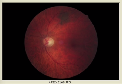
Fundus Photo
It is a retinal photograph used to detect and monitor ocular health. A fundus photograph captures the optic nerve, blood vessels and macula. It can be used to detect glaucoma, macular degeneration, and retinal detachments.
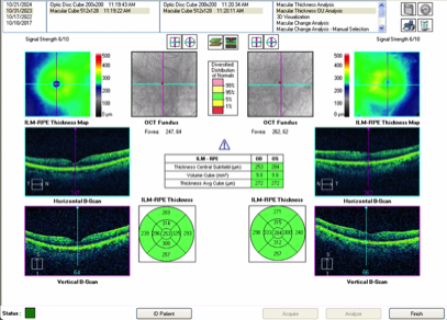
OCT (Optical Coherence Tomagraphy)
Optical Coherence Tomography, advanced technology used to capture internal images of the retina. It is a non-invasive imaging technique used primarily in ophthalmology to capture detailed cross-sectional images of the retina, the light-sensitive tissue at the back of the eye. It’s a vital tool for diagnosing and monitoring various eye conditions, particularly those affecting the retina and optic nerve. OCT works by using light waves to create high-resolution, 3D images of the eye’s internal structures, allowing doctors to see details at a microscopic level.
It can help detect early signs of AMD by visualizing damage or fluid buildup in the macula, the central part of the retina. It also OCT can measure the thickness of the retinal nerve fiber layer, helping detect early signs of glaucoma by assessing optic nerve damage. Additionally, it allows us to see if there is any retinal swelling, fluid leaks, or abnormal blood vessel growth caused by diabetes.

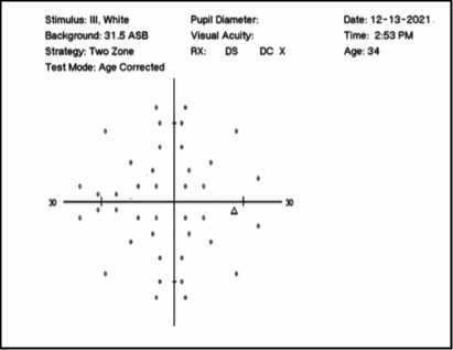
Visual Field
It is a way to measure a patient's central and peripheral vision. It is used to detect any signs of glaucoma, retinal disease and eyelid conditions.
The visual field test can help identify damage to the optic nerve, which may result from various conditions, including optic neuritis, multiple sclerosis, or stroke. Visual field defects caused by optic nerve damage often present as partial vision loss or blind spots, which this test can detect. Additionally, it can reveal visual changes caused by neurological disorders, such as brain tumors, strokes, or pituitary gland tumors. Loss of vision in specific areas (such as half the visual field, known as hemianopia) can indicate damage in particular parts of the brain, helping neurologists identify the affected area and plan appropriate treatment.
-
Retinal photography yields a high-resolution picture of the inside of the eye. This image is kept on file and used as a reference point for future comparisons. This is useful for detection of diabetic changes, hypertensive retinopathy, macular degeneration, optic nerve disease, and retinal holes or thinning.
300 South Beverly Blvd w Suite 307 w Beverly Hills, CA 90212
PROFESSIONAL EYE CARE
(310) 553-2224